
Medical Examinations
Information for health care professionals
Our clinic treats patients with lymphedema based on the medical data that have been accumulated to date. We perform conservative treatment, such as compression therapy, and surgical procedures in an integrated manner, and also focus on building evidence. In cases where intensive conservative treatment is needed, we may refer patients to our affiliated health care providers or clinics. International patients who cannot have tests or examinations in their home country may do them in Japan.
(Note) Please treat DVT (deep vein thrombosis) and cellulitis first, before a patient travels to Japan.
If you have patients with lymphedema, please try to perform as many of the laboratory tests and examinations listed below as possible prior to referral to us. That will help us provide efficient
care to the patients at our clinic. In addition, if any abnormalities are found in these tests or examinations, it will be preferable for you to advise the patient in question to see an
internist, vascular surgeon, and so on prior to visiting us.
【The Importance of Medical Examinations Before Traveling to Japan for Lymphedema Treatment】
If the required examinations cannot be performed in your home country, we will arrange for them to be carried out at our clinic after you arrive in Japan.
However, if these examinations are performed only after your arrival and reveal cardiac abnormalities or significant blood test abnormalities—such as underlying heart disease or internal organ disorders—it may become medically unsafe to proceed with surgery, even for lymphaticovenous anastomosis (LVA) performed under local anesthesia.
For this reason, we strongly recommend that these examinations be completed before traveling to Japan, so that we can confirm in advance that surgery can be performed safely and help ensure a smooth and safe treatment process during your visit.
[ List of tests and examinations ]
(1) Blood test: Blood counts, albumin, liver function, renal function, thyroid gland function, lipids, glucose, coagulability, infection (HIV, syphilis, hepatitis B, hepatitis C, etc.)
(2) Chest X-ray
(3) ECG
(4) Ultrasound examination of leg veins or CT scans of the legs
(5) ABI (Ankle Brachial Index)
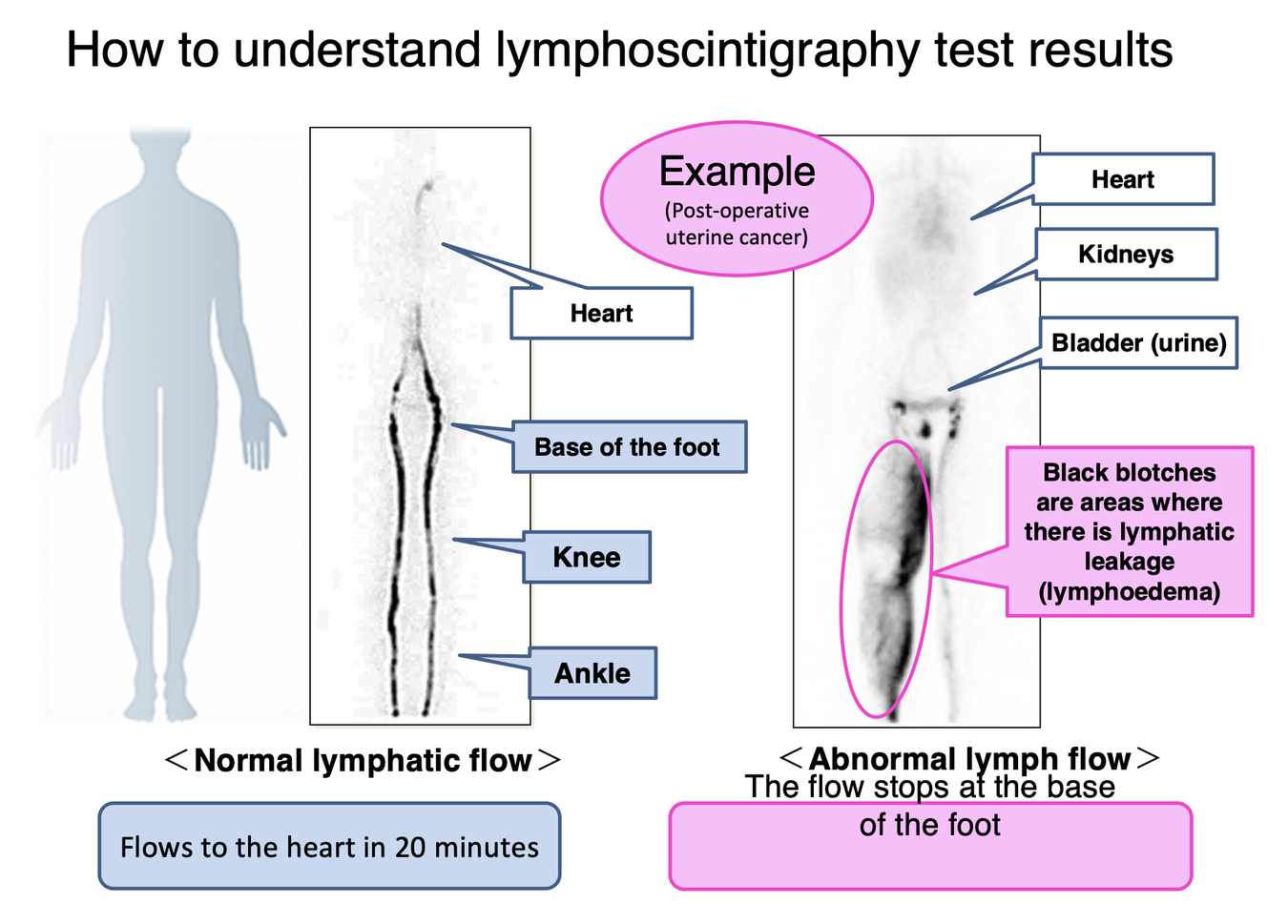
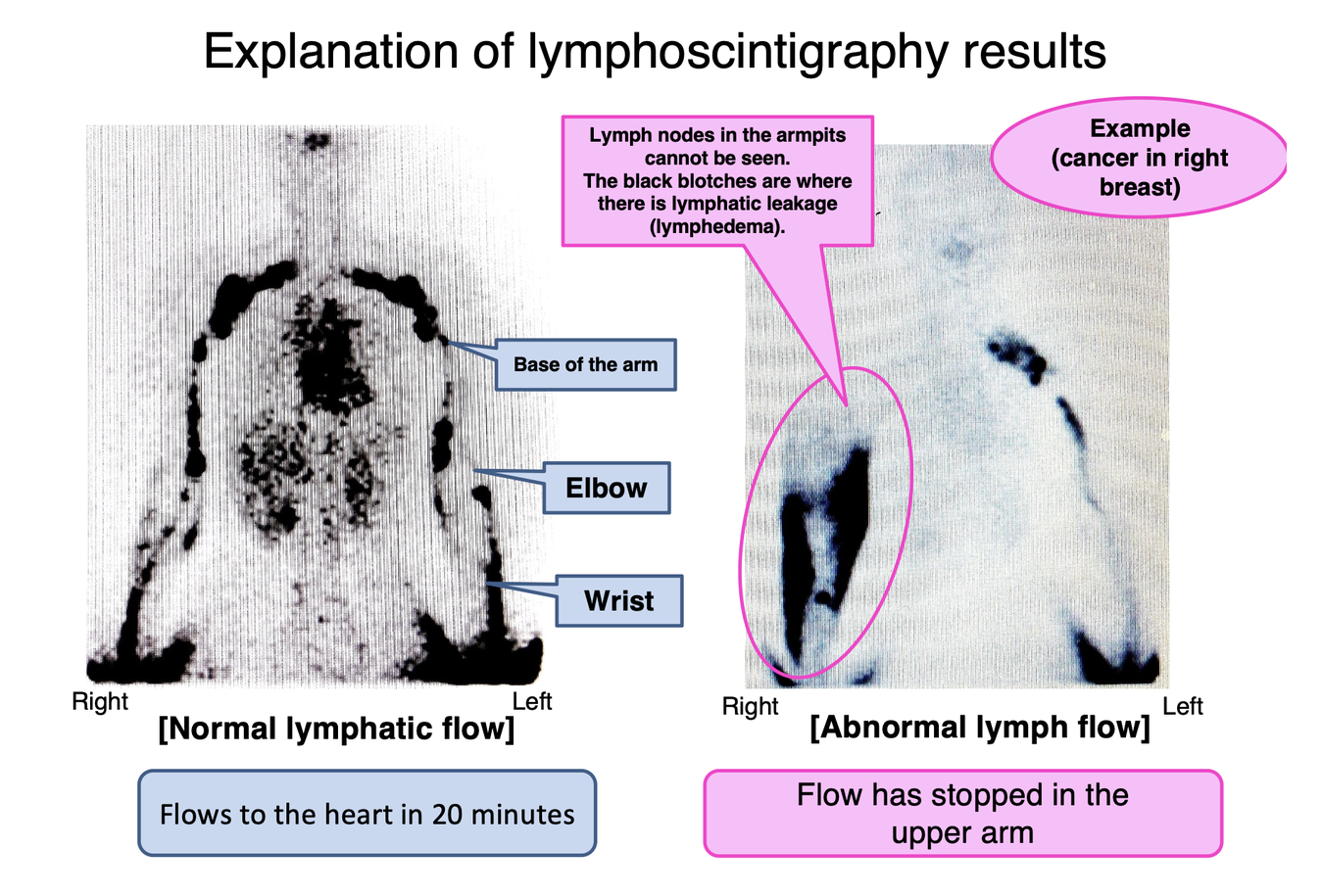
(6) Lymph0scintigraphy
【Examinations Required at the Time of Visit】
Information for Healthcare Professionals
(Referring Physicians / Facilities Performing Examinations)
【Positioning of Examinations at Our Clinic】
At Mukumi Clinic, lymphedema care is conceptualized using a three-layered framework:
1.Confirmation of general systemic condition and differential diagnosis
2.Circulatory evaluation for determining indications for compression therapy and surgery
3.Assessment of lymphatic function
Accordingly, if the examinations listed below can be performed in advance and within feasible limits, the process of clinical evaluation and treatment decision-making will proceed more smoothly.
・If advance examinations are difficult to perform, they will be conducted or scheduled at the time of the patient’s visit to our clinic.
・Explanations of these examinations for patients are provided in the lower section of this page.
【List of Examinations and Their Purposes】
① Blood Tests (Standard Hematology and Biochemical Tests)
Purpose
Screening for hypoalbuminemia, thyroid dysfunction, renal disease, hepatic disease, etc.
→ Exclusion of lymphedema-mimicking conditions (systemic edema)
Evaluation of infection/inflammation and abnormalities in glucose metabolism
Clinical Significance
Differentiation between primary and secondary lymphedema
Safety assessment for conservative treatment and surgical intervention
② Chest X-ray
Purpose
Exclusion of heart failure, pleural effusion, mediastinal pathology, etc.
Clinical Significance
Differential diagnosis of systemic edema and venous return disorders
③ Electrocardiogram (ECG)
Purpose
Evaluation of arrhythmias and ischemic heart disease
Clinical Significance
Safety management for compression therapy and day-surgery procedures
④ Lower Extremity Venous Ultrasound
(Evaluation for DVT and Varicose Veins)
Purpose
Assessment of deep vein thrombosis (DVT) and chronic venous insufficiency
Clinical Significance
Differentiation between lymphedema and venous edema
Reference for initiation and intensity setting of compression therapy
This examination can be performed at our affiliated facility, Ochanomizu Vascular Surgery Clinic.
⑤ ABI (Ankle–Brachial Index)
Purpose
Screening for peripheral arterial disease of the lower extremities
Clinical Significance
Assessment of eligibility and safety for compression therapy
In cases with low ABI values, careful adjustment of compression conditions is required
⑥ Lymphoscintigraphy
Purpose
Global assessment of lymphatic transport capacity
Understanding the pathophysiology of primary versus secondary lymphedema
Differential diagnosis from other internal organ–related diseases
Positioning at Our Clinic
Considered an international standard examination for lymphedema
Used in conjunction with lymphatic ultrasound and ICG lymphography for a comprehensive treatment evaluation
Useful for disease staging, treatment strategy planning, and patient explanation
▶ Recommended Lymphoscintigraphy Protocol at Our Clinic
At our clinic, we routinely perform dual-phase imaging (15 minutes and 60 minutes).
For lower extremities, we emphasize evaluation of physiological lymphatic flow using subcutaneous injections between the toes.
【Recommended Lymphoscintigraphy Protocol】
(Lymphedema Clinic Tokyo)
Equipment and Imaging Settings
Gamma Camera: Symbia Evo Excel
Radiopharmaceutical: 99mTc-HAS-D
Collimator: LMEGP
Matrix Size: 256 × 1024
Zoom: 1.00
Energy Window Preset: 99mTc ±20%, automatic proximity
Detectors: Dual-head detection
Scan Range
Lower extremities: From the ankles to the vertex of the head
Upper extremities: From the wrists to the vertex of the head
Scan Speed
Variable scan speed adjusted so that the total scan time is approximately 10 minutes
Example:
Height 180 cm → 18 cm/min
Height 150 cm → 15 cm/min
Acquisition Phases
15 minutes after injection
60 minutes after injection
After completion of the 15-minute imaging, patients are asked to leave the scanning table and wait until the 60-minute imaging while performing ankle movements, flexion–extension exercises, or gentle massage.
Patient Positioning
Head First
Scan Direction: Head Out
Radiotracer Dose and Injection Method
Dose: 150 MBq per injection
Injection Route: Subcutaneous injection
Local Anesthesia: Lidocaine (Xylocaine) used
Injection Sites
Lower extremities: Subcutaneous injection between the first and second toes
Upper extremities: Subcutaneous injection between the second and third fingers
Clinical Notes
This protocol is designed to evaluate physiological lymphatic flow and overall lymphatic transport function.
It is used as part of a comprehensive lymphedema assessment, in combination with lymphatic ultrasound and ICG lymphography, to support disease staging, treatment planning, and patient education.
【FAQ for Healthcare Professionals】
<Regarding Examinations Required for Clinical Management>
Q1. Why are these examinations reviewed in advance for lymphedema care?
A.
At our clinic, lymphedema is evaluated using the following three-layer approach:
Differential diagnosis (exclusion of systemic and venous edema)
Safety assessment for compression therapy and surgery
Evaluation of lymphatic function
Therefore, having prior information from blood tests, cardiovascular assessments, and vascular evaluations enables us to formulate a concrete treatment strategy from the initial consultation.
Q2. Which blood test parameters are particularly important?
A.
We place particular emphasis on the following:
Albumin (exclusion of malnutrition and systemic edema)
Renal function and hepatic function
Thyroid function
Inflammatory markers
Abnormalities in glucose metabolism
These parameters serve as important indicators for:
Determining whether the condition represents isolated lymphedema
Assessing the safety of conservative therapy and surgery
Q3. Are chest X-ray and ECG required for all patients?
A.
They are not mandatory in all cases; however, they are particularly useful in:
Elderly patients
Patients with a history of cardiac disease
Cases of bilateral or rapidly progressive edema
In patients where heart failure or circulatory insufficiency is involved, lymphedema treatment alone may not lead to improvement, making differential diagnosis crucial.
Q4. How does lower extremity venous ultrasound relate to lymphedema?
A.
In patients with lymphedema, concomitant conditions such as:
Chronic venous insufficiency
Varicose veins
History of DVT
are not uncommon.
Failure to identify these conditions may result in insufficient effects of compression therapy or even symptom exacerbation.
Q5. Why is ABI testing performed?
A.
ABI is regarded as an important tool for evaluating the safety of compression therapy.
Strong compression poses risks in patients with low ABI values
Adjustment of compression pressure and material selection is required
At our clinic, ABI is used not to determine whether compression can be applied, but rather how compression should be applied.
Q6. Is lymphoscintigraphy a mandatory examination?
A.
At our clinic, yes.
If lymphoscintigraphy cannot be performed at your institution, it will be scheduled during the patient’s visit to our outpatient clinic.
Additional examinations may be performed as needed:
Lymphatic ultrasound
ICG lymphography
Lymphoscintigraphy is positioned as a supplementary examination for:
Macroscopic evaluation of lymphatic transport
Understanding the pathophysiology of primary versus secondary lymphedema
Patient explanation and disease staging
Q7. What are the characteristics of the lymphoscintigraphy protocol recommended by your clinic?
A.
At our clinic, we emphasize:
Multi-phase imaging (15, 30, and 60 minutes)
Subcutaneous injection between the toes, which better reflects physiological lymphatic flow
Continuous evaluation from the entire lower limb to the trunk lymphatic flow
For detailed imaging conditions, dosage, patient positioning, and exercise loading, please refer to the separate PDF document titled “Our Recommended Protocol.”
Q8. To what extent do lymphoscintigraphy results influence surgical indication?
A.
They are not used as a sole determinant.
Surgical indications are assessed comprehensively based on:
Lymphatic vessel diameter and wall characteristics on lymphatic ultrasound
Dermal backflow patterns on ICG lymphography
Clinical symptoms and disease stage
Lymphoscintigraphy is positioned as a tool for understanding the overall background of lymphatic flow.
Q9. If examinations have already been performed at another institution, is re-examination necessary?
A.
In principle, no.
If the data are relatively recent (within one year) and clinically consistent, those results will be utilized.
Only missing information will be supplemented as necessary.
Q10. What information would you like us to share at the time of referral?
A.
The following information is particularly helpful:
Primary disease and surgical details (extent of lymph node dissection, history of radiotherapy)
Onset and clinical course of edema
Details of existing compression therapy
History of venous disease and infections (cellulitis)
Results of examinations already performed
This allows us to present concrete treatment options from the initial visit.
Q11. What is the ultimate goal of this examination framework?
A.
The ultimate goals are:
To avoid unnecessary treatments
To avoid unsafe treatments
To present the most appropriate options for each individual patient
At our clinic, our basic policy is to provide care that is not biased toward either conservative therapy or surgery.
Information for Patients with Lymphedema
【How We Approach Lymphedema Care at Our Clinic】
At our clinic, lymphedema is evaluated through a three-layered medical approach:
1.Assessment of your overall health and exclusion of other conditions
2.Circulatory evaluation to determine the safety and appropriateness of compression therapy and surgery
3.Evaluation of lymphatic function
Because of this approach, having certain examinations completed before your visit, whenever possible, allows us to make smoother and more accurate diagnostic and treatment decisions.
If it is difficult to complete these examinations in advance, they can be performed or scheduled after your visit.
A simplified explanation of these tests for patients is provided in the lower section of this page.
【List of Examinations and Their Purpose】
1. Blood Tests (Basic Hematology and Biochemistry)
Purpose
To check for low albumin levels, thyroid dysfunction, kidney disease, or liver disease
To exclude conditions that cause generalized swelling, which may resemble lymphedema
To assess infection, inflammation, and glucose metabolism
Clinical Significance
Differentiation between primary and secondary lymphedema
Safety assessment for conservative treatment and surgical options
2. Chest X-ray
Purpose
To rule out heart failure, pleural effusion, or mediastinal abnormalities
Clinical Significance
Helps distinguish systemic edema or venous circulation problems from lymphedema
3. Electrocardiogram (ECG)
Purpose
Evaluation of arrhythmias or ischemic heart disease
Clinical Significance
Ensures safety when considering compression therapy or day-surgery procedures
4. Lower Limb Venous Ultrasound (DVT / Varicose Veins)
Purpose
Assessment of deep vein thrombosis (DVT) and chronic venous insufficiency
Clinical Significance
Differentiation between lymphedema and venous edema
Helps determine whether compression therapy is appropriate and how strong it should be
This examination can be performed at our affiliated vascular clinic if needed.
5. ABI (Ankle-Brachial Index)
Purpose
Screening for peripheral arterial disease in the lower limbs
Clinical Significance
Safety evaluation before starting compression therapy
In patients with low ABI values, compression must be carefully adjusted
At our clinic, ABI is used not simply to decide whether compression can be applied, but how it should be applied safely.
6. Lymphoscintigraphy
Purpose
Evaluation of overall lymphatic transport function
Understanding the pathophysiology of primary versus secondary lymphedema
Exclusion of other internal medical conditions
【Role at Our Clinic】
Considered an international standard examination for lymphedema
Used in combination with lymphatic ultrasound and ICG lymphography
Helpful for disease staging, treatment planning, and patient explanation
Our Recommended Lymphoscintigraphy Protocol
At our clinic, we typically perform dual-phase imaging (15 minutes and 60 minutes after injection).
For lower-limb lymphedema, we emphasize subcutaneous injection between the toes, as this best reflects physiological lymphatic flow.
【FAQ for Patients】
Q1. Why are these examinations needed before lymphedema treatment?
A.
We evaluate lymphedema from three perspectives:
Excluding other causes of swelling (systemic or venous)
Ensuring the safety of compression therapy and surgery
Understanding lymphatic function
Having blood tests and circulatory evaluations in advance allows us to discuss treatment options more clearly from your first visit.
Q2. Which blood test items are especially important?
A.
We pay particular attention to:
Albumin (nutrition and generalized edema)
Kidney and liver function
Thyroid function
Inflammatory markers
Glucose metabolism
These help us determine whether swelling is caused by lymphedema alone and whether treatment can be performed safely.
Q3. Are chest X-rays and ECGs required for all patients?
A.
They are not mandatory for everyone, but are especially useful for:
Older patients
Patients with a history of heart disease
Patients with bilateral or rapidly progressing swelling
In such cases, swelling may not improve with lymphedema treatment alone, making differentiation important.
Q4. How is venous ultrasound related to lymphedema?
A.
Many patients with lymphedema also have:
Chronic venous insufficiency
Varicose veins
A history of DVT
If these are overlooked, compression therapy may be ineffective or may worsen symptoms.
Q5. Why is ABI testing important?
A.
ABI is essential for evaluating the safety of compression therapy.
Strong compression can be risky in patients with low ABI
Compression pressure and materials must be adjusted carefully
We use ABI to determine how to apply compression safely, not simply whether compression is possible.
Q6. Is lymphoscintigraphy mandatory?
A.
Yes, at our clinic it is considered an important examination.
If it cannot be performed at your referring institution, it will be scheduled during your visit.
Additional tests may include:
Lymphatic ultrasound
ICG lymphography
Lymphoscintigraphy provides a broad overview of lymphatic flow and supports diagnosis and patient education.
Q7. What are the characteristics of your lymphoscintigraphy protocol?
A.
We emphasize:
Multi-phase imaging (15, 30, and 60 minutes)
Subcutaneous injection between the toes to reflect physiological lymph flow
Continuous evaluation from the lower limb to the trunk
Detailed protocols regarding dosage, positioning, and exercise loading are provided separately.
Q8. How much does lymphoscintigraphy influence surgical decisions?
A.
It is not used alone.
We make decisions based on a comprehensive assessment including:
Lymphatic vessel diameter and wall condition on ultrasound
Dermal backflow patterns on ICG lymphography
Clinical symptoms and disease stage
Lymphoscintigraphy provides background understanding of overall lymphatic flow.
Q9. Do examinations need to be repeated if already done elsewhere?
A.
Usually not.
If results are:
Relatively recent (within one year)
Clinically consistent
we will use them. Additional tests are performed only if necessary.
Q10. What information is helpful when being referred?
A.
The following information is very helpful:
Underlying disease and surgical history (including lymph node dissection and radiation)
Onset and progression of swelling
Current compression therapy
History of venous disease or infections (such as cellulitis)
Results of previous examinations
This allows us to discuss treatment options from the very first visit.
Q11. What is the ultimate goal of this examination system?
A.
The goal is to:
Avoid unnecessary treatment
Avoid unsafe treatment
Offer the most appropriate option for each individual patient
Our clinic’s philosophy is to provide balanced care, without favoring either conservative therapy or surgery alone.