Diagnosis
Imaging Evaluation
At our clinic, imaging studies play a central role in the evaluation of lymphedema and in determining the most appropriate treatment strategy.
We combine lymphoscintigraphy, ICG lymphography, and high-resolution ultrasound to assess lymphatic flow, identify areas of dysfunction, and rule out other causes of limb swelling such as deep vein thrombosis (DVT) and venous disease.
By interpreting these studies together, we are able to detect early or subtle lymphatic abnormalities, plan treatment safely, and avoid unnecessary or ineffective procedures.This page explains how imaging guides our clinical decision-making and helps us recommend only the treatment that is appropriate for each individual patient.
【Imaging Studies for Lymphedema】
Early Detection and Treatment Planning Tailored to You
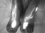
After cancer treatment, some patients notice swelling of the arm or leg.
This condition, known as lymphedema, may gradually worsen if left unaddressed.
Early detection and appropriate evaluation are therefore essential for long-term management.
At our clinic, we combine three complementary imaging studies to accurately assess lymphatic function and to design the most appropriate treatment strategy for each patient.
Each examination provides different information, and together they allow us to recommend only the treatments that are truly necessary and appropriate for you.
【1. Lymphoscintigraphy】
Assessing Lymphatic Flow Throughout the Body
Lymphoscintigraphy is an internationally recognized standard examination for evaluating lymphatic function.
In this test, a very small amount of a radioactive tracer (with established safety) is injected just under the skin of the hand or foot.
Special imaging equipment is then used to observe how lymphatic fluid flows through the body.
This examination allows us to:
Identify areas where lymphatic flow is delayed or obstructed
Evaluate how lymph fluid is drained from the affected limb
Assess the overall lymphatic pathway, including deeper regions of the body
In patients who have undergone lymph node removal during cancer treatment, lymphatic flow may be interrupted, leading to characteristic findings on lymphoscintigraphy.
This examination plays a critical role in determining whether surgical or non-surgical treatment should be considered.
Learn more about lymphoscintigraphy
https://www.english-mominoki-shinryosho.net/diagnosis/lymphoscintigraphy/
【2. ICG Lymphography】
Real-Time Visualization of Superficial Lymphatic Vessels
ICG lymphography uses a fluorescent dye called indocyanine green (ICG), which is injected just beneath the skin.
A near-infrared camera then visualizes lymphatic flow in real time.
This technique allows us to directly observe:
The function of superficial lymphatic vessels
Lymphatic flow patterns close to the skin surface (approximately up to 1 cm depth)
Because ICG lymphography cannot visualize deeper regions—such as the thigh, pelvis, trunk, or areas near the heart—it is always interpreted together with lymphoscintigraphy rather than used alone.
Learn more abut ICG lymphography
https://www.english-mominoki-shinryosho.net/diagnosis/icg-lymphography-indocyanine-green/
【3. High-Resolution Lymphatic Ultrasound 】
Why Lymphatic Ultrasound Matters in Lymphedema Care
Successful lymphedema treatment begins with one essential question:
Which lymphatic vessels remain, where are they, and what condition are they in?
At our clinic, lymphatic ultrasound is not just another imaging test.
It is a core diagnostic step that directly influences treatment decisions and surgical safety.
On our dedicated lymphatic ultrasound page, we explain:
Why lymphatic ultrasound is essential for modern lymphedema care
How it differs from lymphoscintigraphy and ICG lymphography
How precise imaging supports safer, more individualized surgical planning
All explanations are based on clinical experience and scientific evidence, presented in clear and accessible language.
If you have ever felt unsure about:
Whether surgery is truly appropriate for you
Why previous imaging did not provide clear answers
Or whether any options still remain
this page is designed to help you gain clarity and understanding.
Learn more abut lymphatic Ultrasound
【Detecting “Invisible” Lymphedema at an Early Stage】
By selecting and combining these three imaging studies appropriately, we can detect very early changes in lymphatic function—even before noticeable swelling develops.
In some patients, imaging reveals that lymphatic vessels are already severely impaired.
In such cases, LVA surgery may not be the most appropriate option, and alternative treatment strategies are discussed.
Our goal is not to perform surgery whenever possible, but to ensure that the treatment chosen is the most suitable for your condition.
【Supporting Your Future Through Careful Evaluation】
After cancer treatment, many changes occur beneath the surface of the body, even when they are not immediately visible.
For this reason, our clinic emphasizes:
Advanced imaging technology
Careful interpretation by experienced physicians
A multidisciplinary team approach, including specialized therapists, nurses, and allied healthcare professionals
If you are unsure whether your swelling requires treatment, or whether surgery is necessary, we encourage you to consult us.
If imaging shows no significant abnormality, that reassurance itself can be an important and meaningful outcome.
【Imaging Studies for Lymphedema】
Frequently Asked Questions (FAQ)
Q1. Why are imaging studies important for lymphedema?
A.
Lymphedema cannot be evaluated accurately by appearance alone.
Imaging studies allow us to:
Understand how lymphatic fluid is flowing
Identify areas of congestion or dysfunction
Decide whether surgical or non-surgical treatment is appropriate
Our goal is to select the most suitable treatment for each patient, not to perform unnecessary procedures.
Q2. What imaging studies are used at your clinic?
A.
We combine three complementary imaging studies:
Lymphoscintigraphy
ICG lymphography
High-resolution ultrasound examination
Each test provides different information, and no single test is sufficient on its own.
Q3. What is lymphoscintigraphy, and is it safe?
A.
Lymphoscintigraphy is an internationally accepted standard test for evaluating lymphatic function.
A very small amount of radioactive tracer is injected under the skin of the hand or foot.
The radiation dose is low and considered safe for clinical use.
This test allows us to assess:
Whole-body lymphatic flow
Deep lymphatic pathways
Sites of lymphatic obstruction or delay
Q4. What is ICG lymphography, and what can it show?
A.
ICG lymphography uses a fluorescent dye called indocyanine green (ICG) and a near-infrared camera.
It allows real-time visualization of superficial lymphatic vessels, usually up to about 1 cm beneath the skin.
This test is especially useful for:
Evaluating superficial lymphatic function
Planning surgical strategies
However, it cannot evaluate deeper areas such as the pelvis, trunk, or deep thigh regions.
For this reason, it is always interpreted together with lymphoscintigraphy.
Q5. What is the role of ultrasound in lymphedema evaluation?
A.
High-resolution ultrasound allows us to visualize:
Lymphatic vessels beneath the skin
Nearby veins suitable for surgical connection
Surrounding tissue structures
Ultrasound is essential for safe and precise surgical planning, particularly for LVA surgery, as it helps minimize unnecessary incisions and reduce the risk of complications.
Q6. Does ultrasound also check for venous diseases?
A.
Yes.
Lower-limb ultrasound examination includes venous ultrasound to rule out other causes of leg swelling, such as:
Deep vein thrombosis (DVT)
Chronic venous insufficiency
Varicose veins
Lymphedema should be diagnosed only after venous causes of swelling have been excluded.
If swelling is mainly due to venous disease, LVA surgery is not appropriate.
Q7. Can imaging detect lymphedema before visible swelling appears?
A.
Yes.
By combining these imaging studies, we can sometimes detect very early lymphatic dysfunction, even before obvious swelling develops.
Early detection allows:
Timely conservative management
Prevention of progression
Avoidance of unnecessary surgery
Q8. Will imaging always lead to a recommendation for surgery?
A.
No.
Imaging studies are used to guide decision-making, not to justify surgery.
If imaging shows that lymphatic vessels are severely impaired or that swelling is not primarily lymphatic in origin, LVA surgery may not be recommended.
In such cases, alternative treatment strategies are discussed.
Q9. Are these imaging studies used only for surgery planning?
A.
No.
Imaging studies are valuable even when surgery is not planned.
They help:
Confirm or exclude lymphedema
Evaluate disease stage
Optimize conservative treatment strategies
Sometimes, reassurance based on normal imaging findings is itself an important outcome.
Q10. How do these imaging results influence treatment decisions?
A.
Imaging results are interpreted together with:
Clinical symptoms
Physical examination
Lifestyle factors
Based on this comprehensive evaluation, we discuss:
Whether conservative treatment alone is appropriate
Whether surgery may be beneficial
The expected benefits and limitations of each option
Our priority is appropriate patient selection and long-term safety, not procedural volume.
Q11. Do imaging results guarantee treatment outcomes?
A.
No.
Imaging studies help us understand anatomy and function, but they do not guarantee treatment results.
Lymphedema is a chronic condition that requires ongoing management, even after surgical intervention.
Q12. What is your overall approach to imaging and diagnosis?
A.
We use imaging to:
Make lymphedema “visible”
Avoid unnecessary or ineffective treatment
Support informed decision-making
Our philosophy is to recommend only the treatment that is appropriate for your individual condition, based on evidence and careful evaluation.
Patients themselves or their family members are asked to observe the current state of lymphatic vessels while we explain the pathological condition to them. We can select more appropriate treatment methods and predict the effect of treatment by evaluating the lymphatic function.
Ultrasound examinations (upper limb, lower limb, genitalia, lower abdomen)
Conventional lymph scintigraphy and ICG fluorescence lymphography do not make it possible for us to determine the extent of lymphosclerosis, or how much a lymphatic vessel has been damaged. However, the latest technique using ultrasound can allow us to make a diagnosis of lymphosclerosis. We are now more likely than ever to identify better lymphatic vessels.
Learn more about lymphatic ultrasound

Lymphoscintigraphy (upper limb, lower limb, genitalia, lower abdomen)

When radioactive isotopes (drugs that emit low radiation) are injected into the base of the fingers or toes, they are absorbed in the lymphatic vessels. The courses of systemic and deep lymphatic vessels or residual lymphatic function can then be analyzed. With concomitant use of indocyanine green fluorescence lymphography, we can understand the state of the patient's lymphedema in detail and propose the best treatment tailored to the pathological condition. It is possible to appropriately evaluate lymphatic function by taking several images at time intervals after the injections. Even people who are allergic to iodinated contrast media (contrast agents for CT scans) can undergo this scintigraphy.
Lymphoscintigraphy for Lymphedema — Gold Standard Imaging for Accurate Diagnosis and Personalized Treatment Planning in Tokyo
Choosing the right treatment for lymphedema begins with understanding how the lymphatic system is functioning inside the body, not only how much swelling is visible.
Lymphoscintigraphy is internationally recognized as the gold standard imaging test for diagnosing lymphedema.
It provides objective, whole-limb functional information about lymphatic flow, lymph node activity, dermal backflow, and the severity of lymphatic obstruction—insights that cannot be obtained through physical examination alone.
At Lymphedema Clinic Tokyo, we use lymphoscintigraphy as part of a comprehensive, multimodal evaluation, together with ICG lymphography and lymphatic ultrasound, to determine the most appropriate and safest treatment strategy for each patient.
This integrated approach allows us to preserve and maximize remaining lymphatic function and to plan personalized care, including conservative management and lymphaticovenular anastomosis (LVA) when appropriate.
For international patients, we offer online consultations to review lymphoscintigraphy results obtained in your home country.
If lymphoscintigraphy is not available where you live, we can also help arrange the examination at our clinic in Tokyo.
Learn more about lymphoscintigraphy
https://www.english-mominoki-shinryosho.net/diagnosis/lymphoscintigraphy/
What lymphoscintigraphy can (and cannot) show
Why it is essential for accurate diagnosis and staging
How it supports evidence-based, individualized lymphedema care
ICG fluorescence lymphography (upper limb, lower limb, genitalia, lower abdomen)
When a green dye referred to as indocyanine green (ICG) is injected into the base of the fingers or toes, and so on, it is absorbed in the lymphatic vessels. This enables real-time analysis of the courses of superficial lymphatic vessels or residual lymphatic function. This examination is more sensitive than lymph scintigraphy, which makes it possible to reliably diagnose even early lymphedema associated with pain or tightness. It should be noted that people who are allergic to iodinated contrast agents must not undergo this lymphography.
Learn more about ICG lymphography
https://www.english-mominoki-shinryosho.net/diagnosis/icg-lymphography-indocyanine-green/